Top Read Versus Bottom Read for Luminescence
May one, 2012 (Vol. 32, No. 9)
E. J. Dell
Straight Optic Approach Enhances Fluorescent Protein and Other Microplate Analyses
When the Nobel Prize for Chemistry was awarded to Osamu Shimomura, Martin Chalfie, and Roger Y. Tsien for the discovery and development of the green fluorescent protein (GFP) in 2008, researchers around the world were already using GFP (and the many other variants) in a wide range of jail cell-based assays.
One of the well-nigh important uses for GFP is monitoring a diversity of cellular processes in real-time. The many applications that employ fluorescent proteins include: intracellular ship, protein signaling, receptor desensitization, cell movement, migration, sectionalization, apoptosis, metabolism, differentiation, chemotaxis, transcription, translation, and many more.
As with whatever new, popular, Nobel Prize-winning enquiry advancement, a correlative advancement usually occurs in analytical instrumentation. One instrument that all life scientific discipline laboratories have admission to because of the GFP discovery is the confocal microscope. Live, real-fourth dimension pictures (and movies) of cellular processes, highlighted by unlike fluorescent proteins, are hands recorded using a confocal microscope.
Confocal microscopes take immune researchers to obtain fantastic snapshots of biological processes using fluorescent proteins. One drawback of confocal microscopy, though, is that patience and time are needed to obtain reliable, reproducible data since only one cell or cell cluster is viewed at a time.
Some other instrument that has increased in utility due to the GFP discovery is the flow cytometer. More specifically, specialized types of flow cytometers can perform Fluorescence-Activated Cell Sorting (FACS®), which is often used in loftier-throughput screening (HTS) and high-content screening (HCS) labs. FACS provides a way to sort heterogeneous cell populations into homogeneous subgroups, thereby counting and separating the cells that have a fluorescent protein from those that do not.
FACS lack ane of the limitations of confocal microscopy in that it provides an automatic method that counts one specific activated jail cell type. Only similar the confocal microscope, catamenia cytometers that perform FACS are all the same limited past the time information technology can take to obtain data from an experiment with many testable parameters. Hours tin can pass during a FACS experiment, which means that assay conditions may non exist uniform across the unabridged test.
The fluorescent microplate reader is another instrument that has seen an expanded office in life science laboratories after the discovery of GFP. Used mainly in life science or HTS laboratories to study simple, homogeneous absorbance, brilliance, or fluorescence based experiments, microplate readers have evolved into multifunction instruments that can perform circuitous, heterogeneous cell-based assays. Existence able to measure out mL to nL volumes, and upward to thousands of samples at once, microplate readers let for all types of reproducible, jail cell-based assays to be measured in but minutes.
Until recently, though, there was a limitation to cell-based experiments performed in a microplate reader in that the aforementioned sensitivity obtained on a confocal microscope or a FACS could not be matched by a microplate reader. For live, existent-time cell-based experiments, it is preferable to read from the bottom of the microplate.
Reading from the lesser allows for a cover or hat to be placed on top of the microplate to foreclose cell contamination and liquid evaporation. 1 main reason for poorer sensitivity in microplate readers is that they use longer, more than flexible fiber optics to reach the microplate bottom. Since cobweb optics lose light, more signal (and thus sensitivity) is lost when measuring fluorescent proteins in a microplate reader.
To better microplate jail cell-based assays, BMG LABTECH has advanced microplate reader technology past eliminating the need for fiber optics for bottom reading. Using a organization analogous to the microscope, the PHERAstarFS incorporates a series of software-controlled, motor-driven mirrors to focus light through a free air optical path direct onto either the top or bottom of the microplate. Moreover, when switching between superlative and bottom reading modes a unproblematic click in the software is all that is needed, at that place is no irresolute or installation of any additional hardware (i.e., optics, apertures, dichroics, filters, or mirrors). Thus direct-optic bottom reading is fully integrated into the PHERAstarFSoptical system and it completely eliminates the need for fiber eyes.
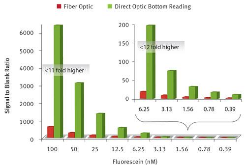
Figure 1. Direct-optic lesser reading (green columns) vs. fiber-optic bottom reading (ruby-red columns): A fluorescein dilution serial in a 1,536-well microplate shows that even at picomolar concentrations the straight-optic bottom reading on the PHERAstar FS will give at least a x times college signal-to-bare ratio than a fiber-optic bottom reading instrument.
Direct-Optic Bottom Reading
To demonstrate the overall comeback of direct-optic lesser reading on the PHERAstarFS as compared to a fiber-optic lesser-reading instrument, a fluorescein dilution series was measured from the bottom of a 1,536-well microplate. As shown inEffigy 1, direct-optic bottom reading gives at to the lowest degree an 11-fold college bespeak-to-blank ratio than fiber-optic bottom reading.
Furthermore, at lower concentrations of fluorescein, the deviation increased to more than 12-fold higher. This will permit for more than 10 times less reagent to exist used on the PHERAstar FS in noncell-based bottom reading assays, significantly saving on reagent costs.
To further demonstrate the overall improvement of directly-optic bottom reading on the PHERAstar FS, GFP-labeled BAE cells (37,500 cells/mL) were measured in a 384-well microplate. As shown in Figure 2, direct-optic bottom reading gives a threefold college signal-to-blank ratio than fiber-optic bottom reading. This will allow for detection of fluorescent proteins at concentrations non possible on any other microplate reader.

Figure 2. Direct-optic lesser reading (greenish column) vs. fiber-optic lesser reading (red column). GFP labeled BAE cells in a 384-well microplate give a three times higher indicate-to-blank ratio when using directly-optic bottom reading on the PHERAstar FS every bit compared to a fiber-optic lesser reading instrument.
With cellular fluorescent protein assays depending sometimes on merely a 25% modify in betoken, a 300% higher bespeak to blank ratio will make experiments possible on a microplate reader that were not possible earlier. This as well means that for secondary cell-based screening assays, fewer cells can be used and thus farther miniaturization is possible on the PHERAstar FS than on other fiber-optic bottom-reading instruments.
In addition to an improved indicate-to-blank ratio in fluorescent protein assays due to direct-optic lesser reading, the PHERAstar FS has a well-scanning fashion that tin create a loftier-resolution epitome of fluorescently labeled cells in each well. Figure 3 shows GFP-tagged HEK293 cells in a 384-well microplate, as visualized using a 20×20 well scanning matrix for each well. This feature enables a digital image to be created from an analog bespeak, which is costless to a microscope or CCD camera that creates analog numbers from a digital picture.

Figure three. Well scanning (xx×20 matrix) and direct-optic lesser reading were used to create a digital epitome of GFP labeled HEK293 cells in a 384-well microplate. Qualitative as well as quantitative data is hands nerveless on the PHERAstar FS.
E.J. Dell, Ph.D. (edward.dell@bmglabtech.com), is the int'fifty marketing manager for BMG LABTECH.
Source: https://www.genengnews.com/magazine/181/bottom-reading-of-cell-based-assays/
0 Response to "Top Read Versus Bottom Read for Luminescence"
Post a Comment